
 Autors: Alberto de Castro, Antonio Benito, Silvestre Manzanera, Juan Mompeán, Belén Cañizares, David Martínez, Jose María Marín, Ireneusz Grulkowski, and Pablo Artal
Journal: Invest Ophthalmol Vis Sci. IOVS
Volume: 59 (nº2)
Pages: 897–903
Year: 2018
ISBN: 1552-5783
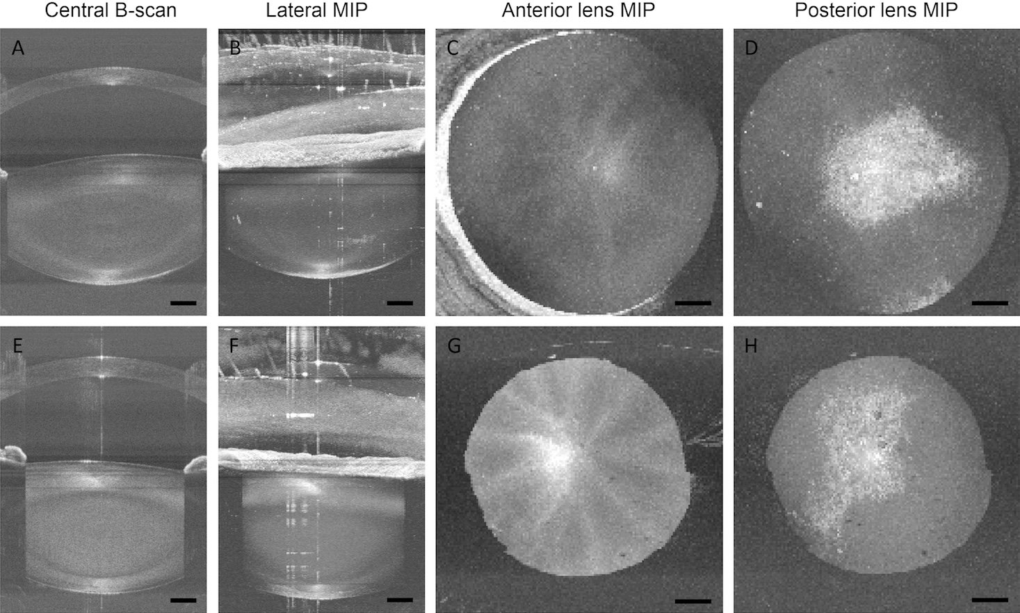
PURPOSE. To image, describe, and characterize different features visible in the crystalline lens of older adults with and without cataract when imaged three-dimensionally with a swept source optical coherence tomography (SS-OCT) system.
METHODS. We used a new SS-OCT laboratory prototype designed to enhance the visualization of the crystalline lens and imaged the entire anterior segment of both eyes in two groups of participants: patients scheduled to undergo cataract surgery, n ¼ 17, age range 36 to 91 years old, and volunteers without visual complains, n ¼ 14, age range 20 to 81 years old. Pre– cataract surgery patients were also clinically graded according to the Lens Opacification Classification System III. The three-dimensional location and shape of the visible opacities were compared with the clinical grading.
RESULTS. Hypo- and hyperreflective features were visible in the lens of all pre–cataract surgery patients and in some of the older adults in the volunteer group. When the clinical examination revealed cortical or subcapsular cataracts, hyperreflective features were visible either in the cortex parallel to the surfaces of the lens or in the posterior pole. Other type of opacities that appeared as hyporeflective localized features were identified in the cortex of the lens. The OCT signal in the nucleus of the crystalline lens correlated with the nuclear cataract clinical grade.
CONCLUSIONS. A dedicated OCT is a useful tool to study in vivo the subtle opacities in the cataractous crystalline lens, revealing its position and size three-dimensionally. The use of these images allows obtaining more detailed information on the age-related changes leading to cataract.
DOI: https://doi.org/10.1167/iovs.17-23596
Download PDF: Three-Dimensional Cataract Crystalline Lens Imaging With Swept-Source Optical Coherence Tomography